- Stroke rates decreased by 77% to 96% among patients undergoing primary cardiac surgery, and by 18% to 100% of patients undergoing redo surgery among those receiving a preoperative CT.
- Mortality decreased by as much as 66% in studies investigating primary surgery.
- CT was used to detect calcifications and to visualize of postoperative anatomy.
- Results of Cochrane systematic review and meta-analysis suggest significantly positive effects of CT scan on cardiac surgery outcomes.
Recent evidence suggests that preoperative computed tomography (CT) imaging (Figure 1) may lead to decreased stroke and mortality rate in patients undergoing cardiac surgery. The use of CT appears to help the surgical team optimize the surgical approach. In patients undergoing redo cardiac surgery stroke rate is also decreased but the effect on mortality is unclear, according to a Cochrane systematic review and meta-analysis published recently in the European Journal of Radiology (http://www.ncbi.nlm.nih.gov/pubmed/26971418)
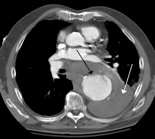
ruptured thoracic aneurysm of about 7 cm.
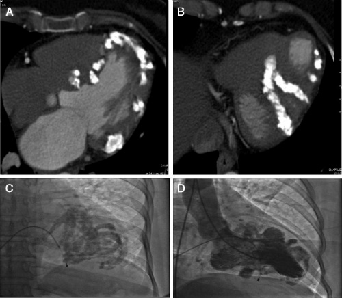
The report, titled “Effect of Computed Tomography Before Cardiac Surgery on Surgical Strategy, Mortality and Stroke” examined whether preoperative chest CT leads to a decrease in postoperative mortality and stroke rate in patients undergoing cardiac surgery. CT was used in these cases to detect of calcifications (Figure 2) and to visualize of postoperative anatomy in redo cardiac surgery, both of which can be used to optimize the surgical approach.
The Analysis
The authors of this report analyzed the PubMed, EMBASE, and Cochrane databases. They searched for articles that examined the use of preoperative CT in cardiac surgery. These were included as were studies concerning primary cardiac surgery and those concerning redo cardiac surgery. Articles not reporting mortality, stroke rate, or change in surgical approach were excluded. (Figure 3.)
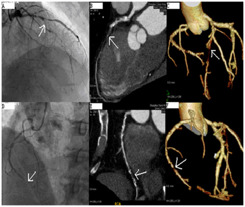
of chronic total occlusion lesions at the left anterior
descending coronary artery (LAD) and right coronary artery (RCA).
The Data
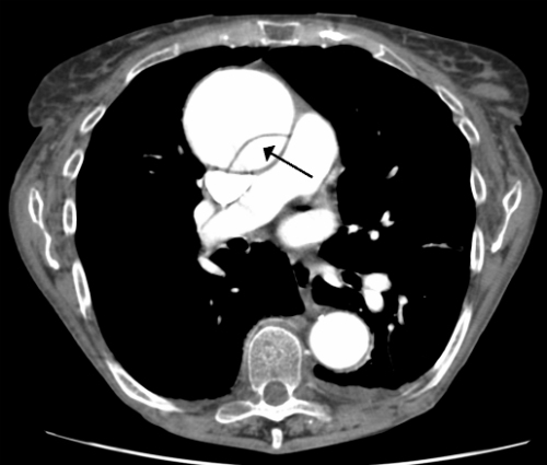
The reviewers found that stroke rates decreased by 77% to 96% among patients undergoing primary cardiac surgery, and by 18% to 100% of patients undergoing redo surgery among those receiving a preoperative CT. Mortality decreased by as much as 66% in studies investigating primary surgery while the effect on mortality in redo surgery varied widely. Change in surgical approach based on CT-findings consisted of choosing a different cannulation site, opting for off-pump surgery and cancellation of surgery. (Figure 4.)
“Current evidence suggests that preoperative CT imaging may lead to decreased stroke and mortality rate in patients undergoing primary cardiac surgery by optimizing surgical approach. In patients undergoing redo cardiac surgery stroke rate is also decreased but the effect on mortality is unclear. However, evidence is weak and included studies were of moderate quality,” the authors concluded.