EGFR Amplification Influences Therapy Response in Patients With Gastroesophageal Cancer
By Cameron Kelsall, /alert Contributor
January 19, 2021
EGFR amplification correlated with poor outcomes in patients with advanced gastroesophageal cancers, with an especially negative prognosis observed in patients treated with EGFR inhibitors combined with chemotherapy, according to an analysis of phase 3 trial data published in Gut.
However, due to the accuracy and feasibility of liquid biopsy testing for EGFR copy number amplifications, clinicians can more easily select appropriate patients for gastroesophageal cancer prospective trials, according to the researchers.
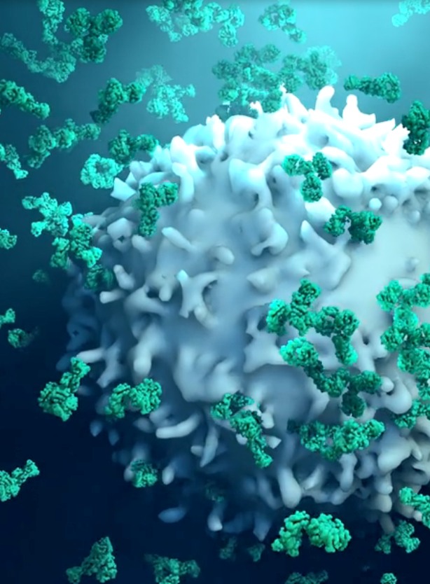
EGFR amplification occurs in 6% to 10% of patients with advanced gastroesophageal cancer. Although several clinical trials failed to show survival improvements among unselected patients treated with cytotoxic chemotherapy and anti-EGFR monoclonal antibodies, emerging data suggest that carefully selected patients may benefit from anti-EGFR therapy.
“If future clinical trials seek to evaluate the efficacy of anti-EGFR therapy in biomarker-enriched populations, understanding the interaction between EGFR amplification and chemotherapy outcome is essential for rational trial design,” wrote Nicola Valeri, MD, PhD, Centre for Molecular Pathology, The Institute of Cancer Research, and colleagues. “Additionally, as heterogeneous expression of biomarkers is a fundamental characteristic of gastroesophageal cancers, analysis of liquid biopsies as a tool to overcome intrapatient heterogeneity is warranted.”
Valeri and colleagues accessed data from patients enrolled in the REAL3 trial, who were assigned to treatment with chemotherapy (epirubicin, oxaliplatin and capecitabine) alone or in combination with panitumumab, an anti-EGFR monoclonal antibody.
In total, 86% of the study’s intent-to-treat population (n = 474 of 553) had EGFR copy number status available by fluorescence in situ hybridization (FISH; n = 114) or digital droplet polymerase chain reaction (ddPCR) in tissue cell-free DNA (n = 250) and plasma cell-free DNA (n = 374).
The researchers sought to determine EGFR detection sensitivity and specificity by screening method. They observed a 98% concordance between FISH and ddPCR. Using FISH as a reference, they reported a ddPCR sensitivity of 1 (95% CI, 0.61-1) and specificity of 0.97 (95% CI, 0.92-0.99); the positive and negative predictive values were 0.81 and 0.95, respectively.
A comparison of tissue and plasma cell-free DNA found an overall concordance of 94.7%. “[W]hen EGFR status was tested in the 60 cases for which data by FISH and ddPCR on tissue, and by ddPCR on cell-free DNA were available, concordance between tissue and blood was 95%,” the researchers wrote.
Compared with patients lacking EGFR amplification, the researchers observed that EGFR amplification (copy number variation score ≥2) was associated with shorter median progression-free survival (PFS; 6.4 months vs. 4.6 months) and overall survival (OS; 11.2 months vs. 9.7 months) in the intent-to-treat population.
The lack of benefit persisted when the researchers employed a higher threshold (≥5) to the copy number variation score (hazard ratio for PFS, 1.7; hazard ratio for OS, 1.5).
In order to validate their findings in a larger cohort, the researchers accessed the cBioportal for Cancer Genomics, identifying 2122 EGFR samples from 2054 treated in 11 discrete studies.
“In line with our findings, EGFR amplification was observed in approximately 6.5% of patients and was consistently associated with inferior OS, disease-specific survival, disease-free survival and PFS in the validation cohorts,” Valeri and colleagues wrote. “Taken together these data strengthen the notion that EGFR amplification may be a prognostic biomarker in gastroesophageal adenocarcinoma.”
Poor outcomes persisted in a cohort of patients with ddPCR cell-free DNA data, in which patients with EGFR amplification consistently had worse PFS and OS than those without amplification.
The researchers further observed that treatment with panitumumab did not improve outcomes in patients with EGFR amplification; conversely, it appeared to provoke worse outcomes. “Despite the observation that adding panitumumab had a negative prognostic role in both groups, it was unexpected to notice that the negative association between EGFR inhibitors and [chemotherapy] appeared more pronounced in patients whose tumour harboured an EGFR amplification,” they wrote.
Patient-derived organoids (PDOs) were developed to evaluate the relationship between anti-EGFR agents and chemotherapy in patients with EGFR amplification. The researchers observed a negative interaction between the chemotherapeutic agent epirubicin and anti-EGFR therapy in EGFR-amplified PDOs.
“Our observations suggest that inhibition of EGFR signalling in a background of epirubicin treatment selectively induces the downregulation of the G1/S negative regulator p21 and the upregulation of the S phase-promoting cyclin E1 in EGFR-amplified gastric cancer PDOs, thereby leading to increased flux to the S phase of the cell cycle,” the researchers wrote. “Cyclin E1 upregulation has been linked to shortened mitosis and increased mitotic exit.”
Potential study limitations identified by the researchers included the small sample size and the lack of consistent chemotherapy dosing in different arms of the REAL3 trial.
“Our results also emphasize the challenge in designing optimal drug combinations in absence of robust patient-centred preclinical models,” the researchers concluded. “In view of the relatively rarity of EGFR-amplified gastroesophageal cancers, we suggest that international collaborative efforts may be required in order to facilitate prospective trials enriched for EGFR-amplified tumors based on liquid biopsy testing.”
MORE FROMGastric Cancer Advanced Learning Center


.jpg)

Atezolizumab, Bevacizumab Combination Boosts Survival for Liver Cancer
February 10, 2021
Triplet Therapy Shrinks HER2-Positive Gastric Cancer Tumors
February 05, 2021


Investigational BRAF-Inhibitor Demonstrates Benefit in Early Trials
January 19, 2021

Surgeon Technical Skill Has Big Impact On Colon-cancer Outcomes
January 19, 2021

Gastric Cancer Predicted to Become a Rare Disease
January 19, 2021